
|
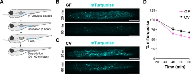
Fig. 2 Lysosome-rich enterocyte (LRE) protein degradation activity is reduced by microbes. (A) Cartoon depicting the experimental design of pulse-chase protein uptake and degradation assay. At 6 dpf, germ-free (GF) and conventional (CV) larvae were gavaged with mTurquoise (25 mg/mL), incubated for 1 hr, and then flushed with PBS to remove luminal mTurquoise. LRE degradation of mTurquoise was measured by confocal microscopy over time. (B, C) Confocal images of mTurquoise fluorescence in the LRE region after flushing in GF (B) and CV (C) larvae (scale bars = 50 µm). (D) Plot showing the degradation of mTurquoise fluorescence (%) in the LRE region over time. Degradation occurred at a significantly faster rate in GF than CV larvae from 20 to 60 min post gavage (Simple linear regression, p=0.0167, n=6).