
|
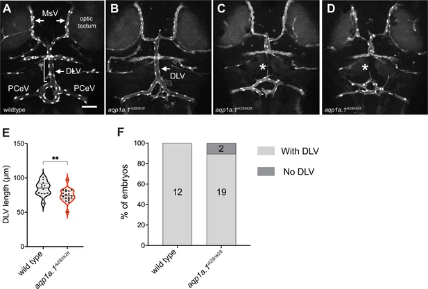
Fig. 3 - Supplemental 6 Cerebral vascular formation defects in aqp1a.1 mutant zebrafish. (A) Dorsal view of 4 dpf wildtype cranial vasculature visualized by Tg(fli1a:H2B-EGFP)ncv69;(fli1:Lifeact-mCherry)ncv7 expression, showing distinct cranial vessels. (B–D) Dorsal view of cranial vasculature of 4 dpf aqp1a.1rk28/rk28 zebrafish, showing one representative embryo with DLV (B, arrow) and two other embryos lacking the DLV (C and D, asterisks). (E) Quantification of DLV lengths in embryos that formed the DLV at 4 dpf (n = 12 wildtype embryos; n = 19 aqp1a.1rk28/rk28 embryos, two independent experiments). Statistical significance was determined by unpaired t-test; **p<0.01. (F) Percentage of the embryos of indicated genotype with and without the DLV at 4 dpf (n = 12 wildtype embryos, n = 21 aqp1a.1rk28/rk28 embryos, two independent experiments). DLV, dorsal longitudinal vein; MsV, mesencephalic vein; PCeV, posterior cerebral vein. Scale bar, 50 µm.