
|
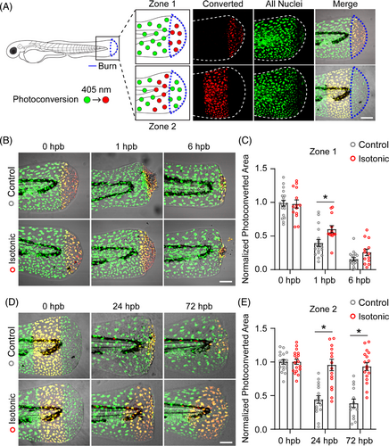
Fig. 2 Keratinocyte motility contributes to spread of epithelial damage after burn. (A) Schematic of photoconversion experiment. Keratinocyte nuclei expressing Dendra2 (green) can be photoconverted (red) by exposure to 405-nm light. Photoconversion was performed in the indicated region immediately following burn injury indicated by the blue outline. In merged images, photoconverted nuclei appear yellow due to residual green Dendra2 fluorescence. (B) Images showing control and isotonic medium treated larvae after photoconversion of zone 1 tissue. (C) Quantification of zone 1 photoconverted area at the indicated time, hours post-burn (hpb). Data are normalised to photoconverted area 0 hpb. N ≥ 18 control and N ≥ 13 isotonic treated larvae. (D) Images showing control and isotonic medium treated larvae after photoconversion of zone 2 tissue. (E) Quantification of zone 2 photoconverted area at the indicated time, hours post-burn (hpb). Data are normalised to photoconverted area 0 hpb. N ≥ 14 control and N ≥ 16 isotonic treated larvae. Scale bars = 100 μm. Asterisk indicates p < 0.05 by two-way ANOVA.