
|
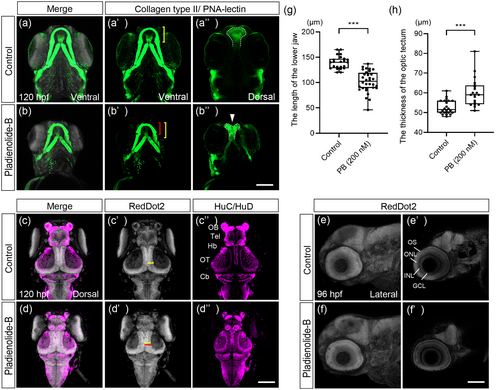
Fig. 6 Craniofacial malformation and structural abnormalities of the brain and eye in PB-exposed embryos at 96 and 120 hpf. (a–b″) Immunofluorescence images of the control and PB-treated (200 nM) embryos. Samples were stained with anti-collagen type II antibody and PNA-lectin to visualize facial cartilages. The Meckel's cartilage ((a′), (b′), ventral view) and ethmoid plate ((a″), (b″), dorsal view) indicated by white arrowhead were analyzed. The bracket indicates the length of the Meckel's cartilage in control (yellow bracket) and PB-treated embryos (red bracket), while dashed lines outline the ethmoid plate. (c–d″) Samples were stained with anti-HuC/HuD and RedDot2 to analyze brain defects. Bracket shows the thickness of optic tectum in control (yellow bracket) and PB-treated embryos (red bracket). (e–f′) Eye morphology ((e), (f)) and eye layer ((e′), (f′)) were analyzed. (g) Quantification of the length of the lower jaw shown in panels (a′) and (b′). (h) Quantification of the thickness of the optic tectum shown in panels (c′) and (d′). ***: p < 0.001 (one-way ANOVA followed by Dunnett's multiple comparison test). Cb, cerebellum; GCL, ganglion cell layer; Hb, habenula; INL, inner nuclear layer; OB, olfactory bulb; ONL, outer nuclear layer; OS, outer segment of photoreceptor; OT, optic tectum; Tel, telencephalon. Scale bars: 100 μm. n = 19, 28, 21, and 28 for experiment for (a, c), (b, d), (e), and (f), respectively. The dots in (g) and (h) represent individual embryos examined. All experiments were performed in three biological replicates.