
Image
|
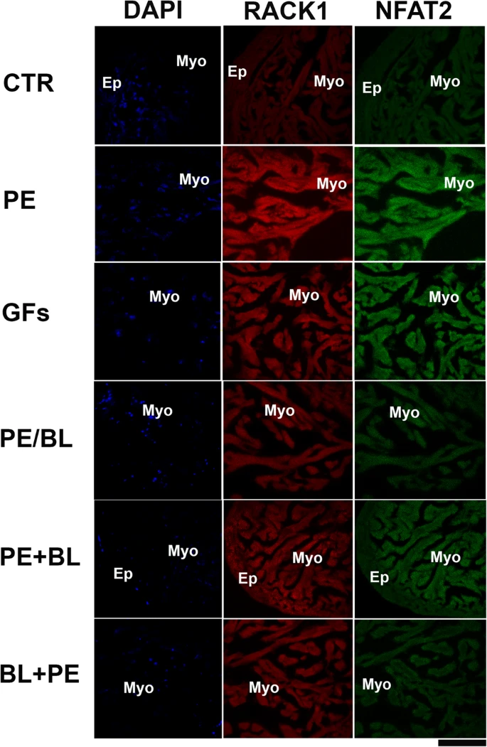
Figure Caption
Fig. 5 Double staining of RACK1 and NFAT2 was performed via confocal microscopy. RACK1 (red fluorescence) is fundamentally expressed in the myocardium of CTR. In all the experimental groups, RACK1 expression was increased, and RACK1 colocalized with the NFAT2 antibody to mark the endocardium (green fluorescence). NFAT2 is strongly expressed in PE and GFs and moderately expressed in PE + BL, PE/BL, and BL + PE. The hearts utilized for the experiments were N = 4/5 sections in each group in triplicate experiments. Blue fluorescence: DAPI (nuclear marker); scale bar: 500 μm.
Acknowledgments
This image is the copyrighted work of the attributed author or publisher, and
ZFIN has permission only to display this image to its users.
Additional permissions should be obtained from the applicable author or publisher of the image.
Full text @ Sci. Rep.