
|
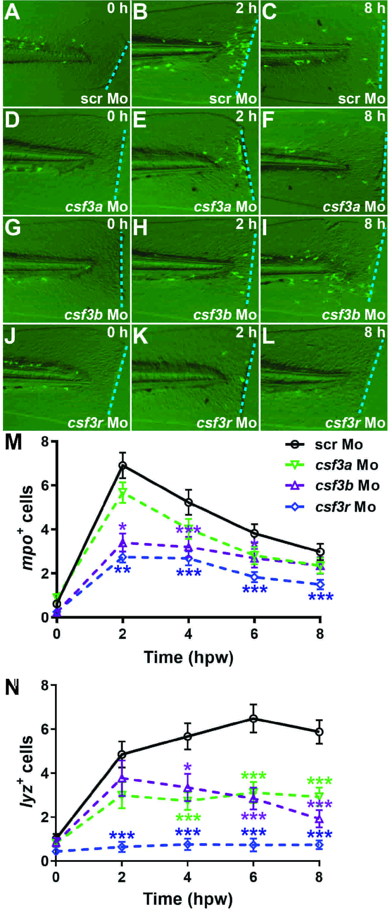
Fig. 2
Role of alternative G-CSFR ligands in response to wounding. (A–L) Fluorescent microscopy images of representative Tg (mpo::GFP) embryos injected with either scr Mo (A–C), csf3a Mo (D–F), csf3b (G–I) or csf3r Mo (J–L) at the indicated times post wounding. (M,N) Quantitation of recruitment of mpo+ (M) and lyz+ (N) cells to the wounding site at 0, 2, 4, 6 and 8 hours post wounding (hpw) in Tg (mpo::GFP) and Tg (lyz::DsRed) embryos, respectively, that had been injected with the indicated morpholinos, showing mean and S.E.M. along with statistical significance of gene-specific in comparison to scr Mo (*: p