IMAGE

Fig. 3

ID
ZDB-IMAGE-210424-42
Source
Figures for Brunt et al., 2021
Image
Figure Caption

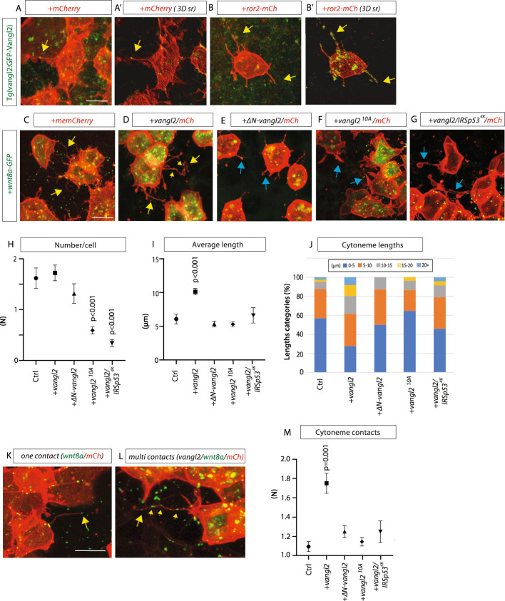
Fig. 3 Vangl2 regulates length and contacts of Wnt8a cytonemes in the zebrafish embryo.

A, BTg(vangl2:GFP-Vangl2) zebrafish embryos were injected with mem-mCherry (mCh) or Ror2-Mcherry. A’, B’ 3D renders (3D sr) of A&B at 5hpf. Yellow arrows show examples of Vangl2 positive cytonemes. AAn = 6 embryos. BB’: n = 8 embryos. CG Wild type zebrafish embryos were injected with indicated constructs and imaged at 5hpf. Yellow arrows show examples of Wnt8a positive cytonemes. Blue arrows show examples of thick membrane protrusions. Scale bar = 10 µm. H Number of Wnt8a positive cytonemes per cell (N = number). (n per condition = 3, 6, 3, 9, 7 embryos, n = 87, 123, 44, 154, 93 cells). I Length of Wnt8a positive cytonemes (µm). (n per condition = 81, 269, 54, 82, 24 cytonemes). J Breakdown of the percentage of Wnt8a positive cytoneme lengths into 0–5, 5–10, 10–15, 20+ µm categories. K Wnt8a/Mem-mCh cytoneme with one wnt8a + ve contact point. Scale bar = 10 µm. L Vangl2/Wnt8a/mem-mCh cytoneme with multiple contact points M Quantification of number of Wnt8a-positive contact points per cytoneme. Wnt8a positive points were counted when at tips or at a fixed junction or “turning point” (n = 32, 125, 80, 81, 24 cytonemes). Graphs represent mean and standard error of the mean. Corresponding dot plots are shown in Supplementary Fig. 5. H, I, K Two-sided Kruskal–Wallis tests with Bonferroni correction for multiple tests. Statistical significance: p ≤ 0.05. SEM = 1. Source data are provided as a Source Data file.

Acknowledgments
This image is the copyrighted work of the attributed author or publisher, and ZFIN has permission only to display this image to its users. Additional permissions should be obtained from the applicable author or publisher of the image. Full text @ Nat. Commun.