
Figure 4
- ID
- ZDB-IMAGE-201229-67
- Genes
- Publication
- Keefe et al., 2020 - Vanishing white matter disease expression of truncated EIF2B5 activates induced stress response
- All Figures
- Figures for Keefe et al., 2020
|
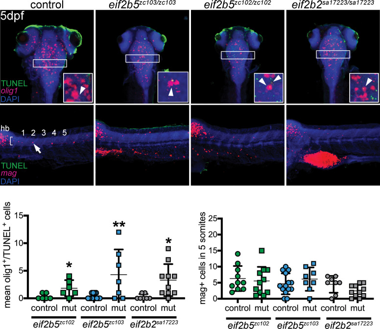
Figure 4 Top row (and quantification in lower left): increased apoptotic OPCs in the brain and spinal cord. Confocal images of brain, z-stack, rostral to the top, double-labeling for TUNEL and olig1 (in situ probe), in WT, eif2b5zc103/zc103, eif2b5zc102/zc102 or eif2b2sa17223/sa172233 larvae. Region used for quantification shown by box. Inset in each panel shows example of double-labeled cell (TUNEL, olig1) for each genotype (except WT), single confocal slice image. Middle panels: no change in apoptosis of differentiated oligodendrocytes, co-labeled with myelin associated glycoprotein (mag) (in situ probe) and TUNEL. Confocal z-stack images of spinal cord, rostral to the left; quantified in lower right panel.