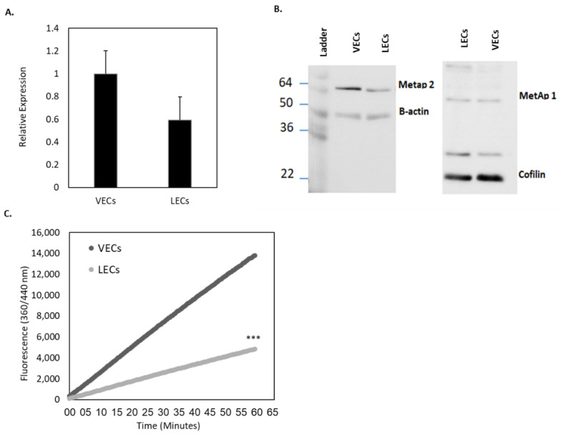
IMAGE

Figure 2

ID
ZDB-IMAGE-200814-5
Source
Figures for Esa et al., 2020
Image
Figure Caption

Figure 2

Basal MetAp2 activity and expression in LECs compared with VECs. (A) The MetAp2 mRNA levels in LECs (HMVEC-dLyAd cells) is ~60% of the mRNA levels in VECs (HUVECs), as measured by qRT-PCR (5 μg), n = 3. (B) Western blot analyses for determining the expression of MetAp2 in both cell lines. (C) Measurement of the basal activity of MetAp2 in HUVECs and HMVEC-dLyAd cells. A homogenate containing an equal number of cells was added to an L-Met-AMC solution (250 µM). The reaction was monitored for 1 h via the fluorescent quantification of the cleaved substrate. MetAp2 in HMVEC-dLyAd cells showed over a 65% reduction in the enzymatic ability to cleave labeled methionine when compared with HUVECs. The results were normalized to the total cell number. *** p < 0.001. The results are presented as mean ± SEM.

Acknowledgments
This image is the copyrighted work of the attributed author or publisher, and ZFIN has permission only to display this image to its users. Additional permissions should be obtained from the applicable author or publisher of the image. Full text @ Int. J. Mol. Sci.