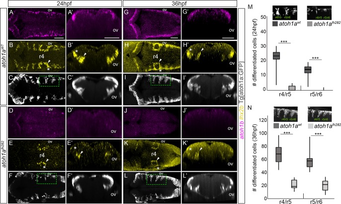
atoh1a is required for the specification of the lhx2b neuronal population.
A-L) atoh1aWT and atoh1afh282 embryos in the Tg[atoh1a:GFP] background were analyzed at 24hpf (atoh1aWT n = 14; atoh1afh282 n = 18) and 36hpf (atoh1aWT n = 11; atoh1afh282 n = 7) with atoh1b (A, D, G, J), lhx2b (B, E, H, K), and anti-GFP in order to follow the atoh1a-derivatives (C, F, I, L). A’-L’) Reconstructed transverse views of dorsal views displayed in (A-L) at the level of the anterior side of the otic vesicle. Note that atoh1b expression (compare A-A’ and G-G’ with D-D’ and J-J’), and the lateral domains of lhx2b diminished (compare white asterisks in B-B’ with E-E’, and H-H’ with K-K’), whereas the more medial domain does not decrease so dramatically (compare white arrowheads in B-B’ with E-E’, and H-H’ with K-K’). Note that atoh1a:GFP cells remained, suggesting that there is no massive cell death. M-N) Quantification of differentiated neurons in the r4/r5 and r5/r6 domains of atoh1aWT and atoh1afh282 embryos as depicted in the small inserts showing dorsal views of halves hindbrains that correspond to the framed regions in (F-L), *** p<0.001 (Table 1 for values and statistical analysis). Note the reduction in the number of atoh1a:GFP differentiated neurons in atoh1afh282 embryos. ov, otic vesicle; r, rhombomere. Scale bars correspond to 50 μm.
This image is the copyrighted work of the attributed author or publisher, and
ZFIN has permission only to display this image to its users.
Additional permissions should be obtained from the applicable author or publisher of the image.
Full text @ PLoS One
Your Input Welcome
Thank you for submitting comments. Your input has been emailed to ZFIN curators who may contact you if
additional information is required.
Oops. Something went wrong. Please try again later.