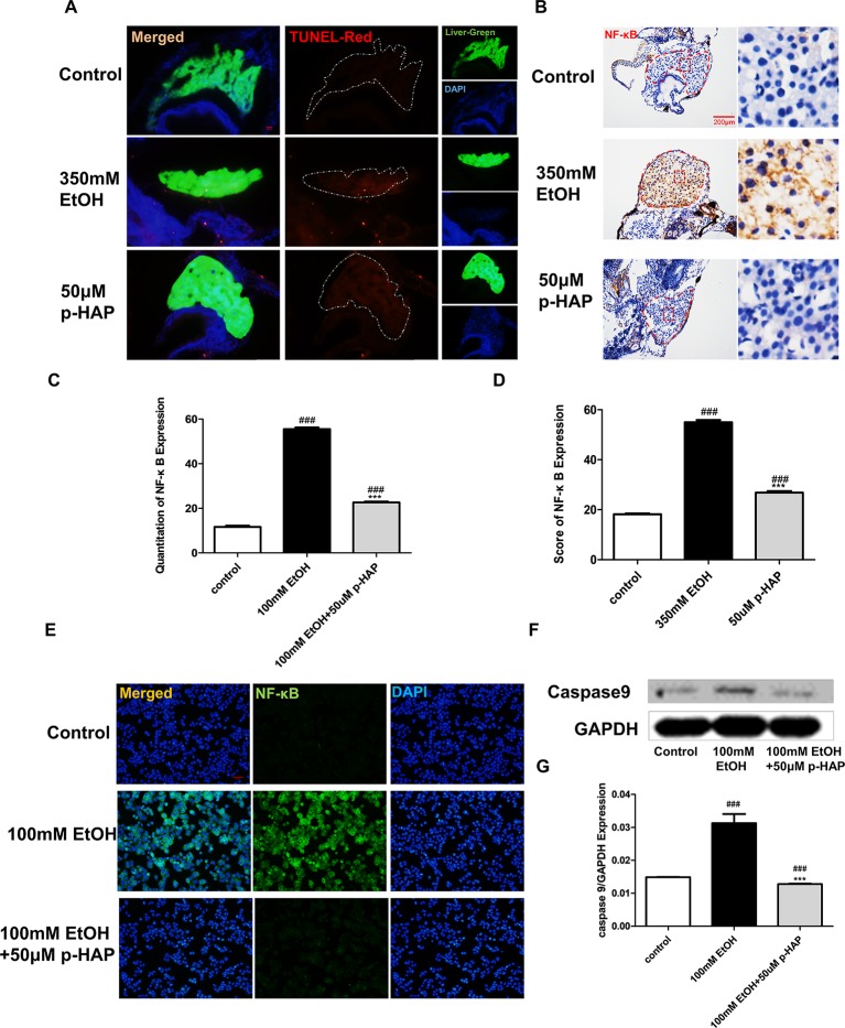
P-HAP Protected Zebrafish Larvae Against Apoptosis and Reduced Inflammation After Alcohol Administration. (A)In situ detection of cell apoptosis by TUNEL staining of paraffin liver sections in control zebrafish larvae and zebrafish larvae treated with 350 mM ethanol or 50 μM p-HAP. Apoptotic cells are indicated by white arrowheads. Figures are magnified as 400×. (B) NF-κB immunohistochemical staining of zebrafish larvae. Figures are magnified as 400×. (C) Quantitative analysis of NF-κB expression. (D) The amounts of NF-κB expression were quantified according to fluorescence intensity. (E) NF-κB immunofluorescence staining of LO2 slides. Figures are magnified as 200×. (F) Western blot analysis of caspase-9 expression in LO2 treated with 350 mM ethanol and 50 μM p-HAP. (G) Quantitative analysis of caspase-9 protein expression. Data are expressed as the mean ± SD. ###P < 0.0001 vs control group. ***P < 0.001 vs model group.
Acknowledgments
This image is the copyrighted work of the attributed author or publisher, and
ZFIN has permission only to display this image to its users.
Additional permissions should be obtained from the applicable author or publisher of the image.
Full text @ Front Pharmacol
Your Input Welcome
Thank you for submitting comments. Your input has been emailed to ZFIN curators who may contact you if
additional information is required.
Oops. Something went wrong. Please try again later.