IMAGE

FIGURE 3

ID
ZDB-IMAGE-191230-66
Source
Figures for König et al., 2019
Image
Figure Caption

FIGURE 3

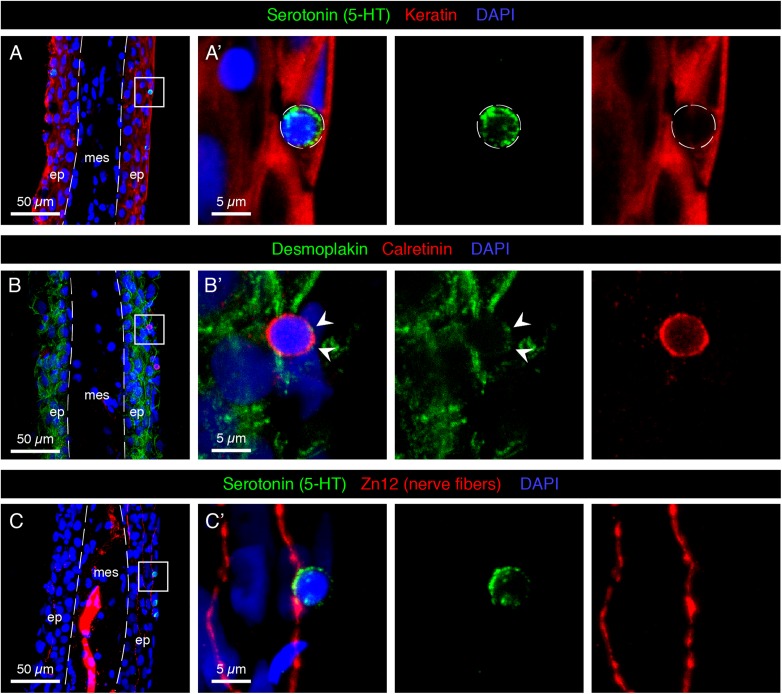
Characterization of HCS-cells in the adult uninjured fin. (A–C) Immunofluorescence staining of longitudinal sections of uninjured adult caudal fins; ep, epidermis, mes, mesenchyme. Dashed lines indicate the border between epidermis and mesenchyme. (A,A’) Serotonin-positive cells (green) do not express Keratin (red), which demarcates surrounding epidermal cells. (B,B’) Immunostaining for Desmoplakin (green) and Calretinin (red) indicates that Calretinin-positive cells possess very few desmosomes compared to the surrounding keratinocytes. Very weak dotty Desmoplakin staining is sometimes observed at the apical side of the cell (arrowheads). (C,C’) Staining for Serotonin (green) and Zn12 (red), a neuronal marker, reveals that cells are located close to nerve fibers. N ≥ 4 for each staining. Nuclei are labeled with DAPI (blue).

Acknowledgments
This image is the copyrighted work of the attributed author or publisher, and ZFIN has permission only to display this image to its users. Additional permissions should be obtained from the applicable author or publisher of the image. Full text @ Front. Mol. Neurosci.