
Fig. 4
- ID
- ZDB-IMAGE-190816-31
- Antibodies
- Publication
- Fogerty et al., 2019 - Genomic non-redundancy of the mir-183/96/182 cluster and its requirement for hair cell maintenance
- All Figures
- Figures for Fogerty et al., 2019
|
Fig. 4
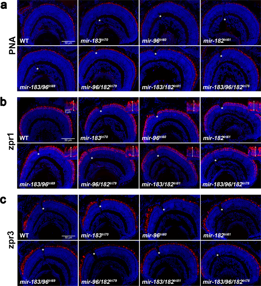
Survey of retinal anatomy in 5dpf mutant larvae. Transverse sections were collected near the optic nerve, which is visible in some images (asterisks), and stained with markers for different photoreceptor cell types. In all images, the ventral retina is to the left. (a) Peanut agglutinin lectin (PNA) labels the extracellular matrix surrounding cone outer segments. (b) zpr1 labels red/green cone inner segments. Inserts show higher magnification images, with double arrows indicating how inner segments were measured. (c) zpr3 labels rod outer segments, which are immature at this age and typically longer and more numerous in the ventral retina.