
|
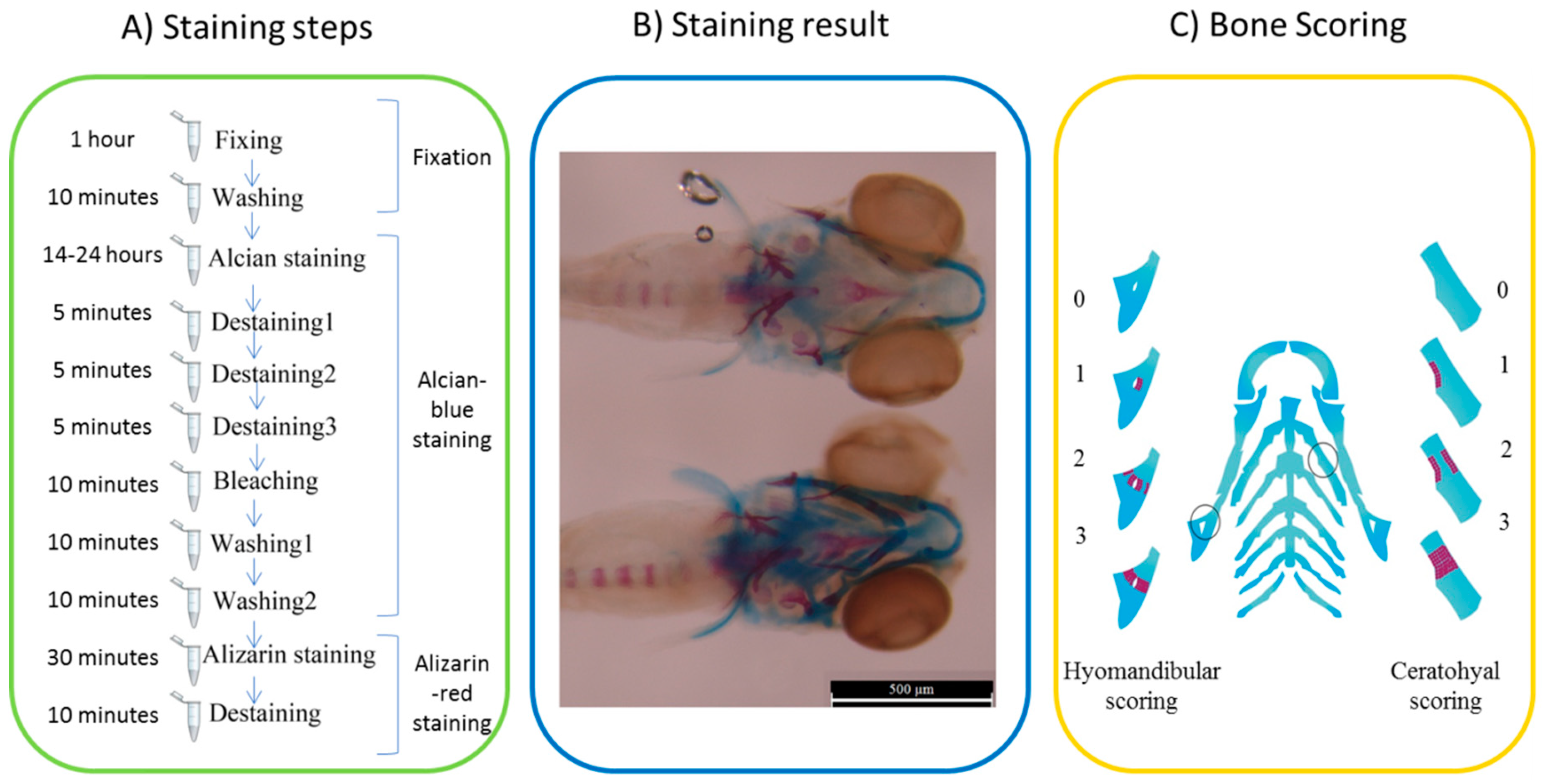
Fig. 8
Two color staining and scoring system of the target bones in treated zebrafish larvae at 6 dpf. (A) Alcian-blue and alizarin-red staining steps, (B) sample of the stained ferutinin-treated larvae: blue parts represent ceratohyal and red parts demonstrate mineralized sections and the scale bar is 500 μM, and (C) ceratohyal and hyomandibular scoring system: 0 when the target area was just blue without bone mineralization, 1 for bone mineralization of one part of target cartilage, 2 for red stained two parts of cartilage but not completed, and 3 was for completely mineralized bone. Circles showed the position of the ceratohyal and hyomandibular as target bones.