
|
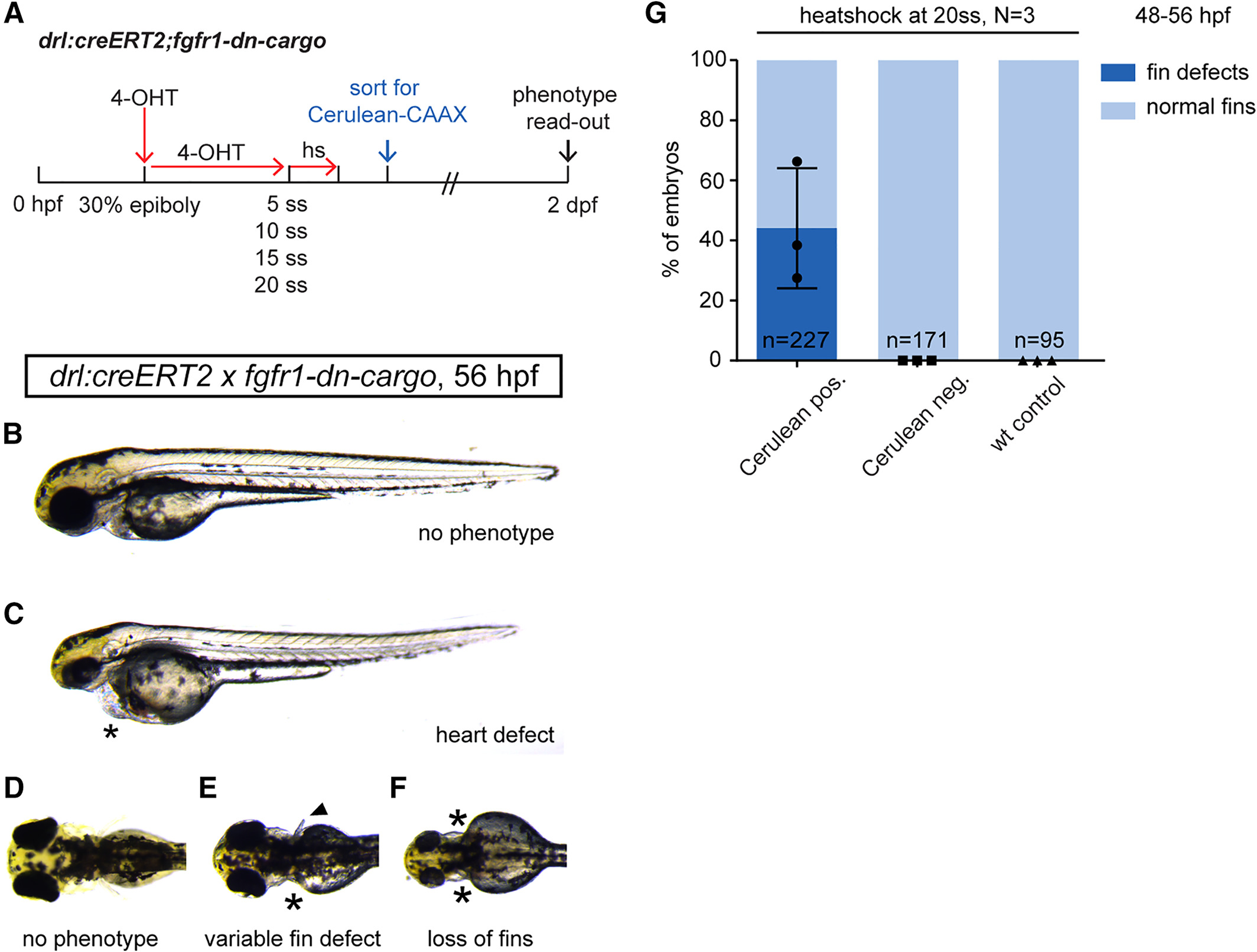
Fig. 7
Temporal dissection of tissue‐specific FGF signaling control of cardiac and fin development. A: Schematic showing experimental time line of tissue‐specific FGF signaling perturbations at discrete developmental stages. 4‐OHT induction at 30% epiboly triggers Cre/lox recombination in earliest drl‐expressing progenitors. Heatshock treatments at distinct time points throughout somitogenesis target different phases of cardiac and fin development. B–D: Phenotypes scored in 2‐dpf drl:creERT2;fgfr1‐dn‐cargo embryos treated as indicated in the schematic above. B,C: Heart defects as scored through blood pooling and cardiac edema on top of the yolk (asterisks); lateral views, anterior to the top. D–F: Fin defects were counted upon unilateral; or bilateral fin deformations (arrow head in E, compare to normal pectoral fins in D) and/or loss (asterisks in E,F). G: Quantifications of phenotypes in drl:creERT2;fgfr1‐dn‐cargo double transgenics heatshock‐treated at different time points during somitogenesis. LPM‐specific FGF perturbation triggered by heatshock at 20 ss results in pectoral fin defects in on average 44.09% (s.d. 20.01, p = 005, total n = 227, three independent experiments) of double‐transgenic embryos with prior Cerulean expression; earlier heatshock timings caused no discernible fin defects. Number “n” indicates the number of individual embryos analyzed per condition; N indicates the number of individual experiments performed. Statistics based on one‐way ANOVA, multiple‐comparison Tukey's post‐test.