
Image
|
Figure Caption
Fig. S1
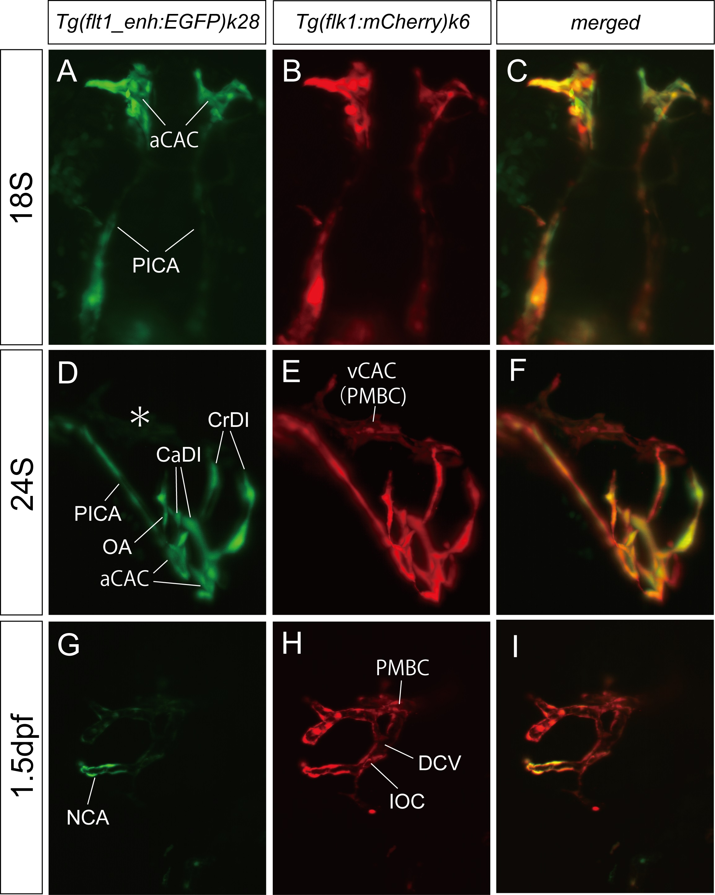
Arterial and venous characteristics of each ocular vessel.
Light-sheet microscopy of the double Tg(flt1_enh:EGFP)k28 and Tg(flk1:mCherry)k6 zebrafish embryos at 18S (A-C), 24S (D-F), and 1.5 dpf (G-I). Tg(flt1_enh:EGFP)k28 (A, D, and G), Tg(flk1:mCherry)k6 (B, E, and H), and merged (C, F, and I) images. Dorsal (A-C), rostral-lateral (D-F), and lateral (G-I) views. Only the arterial components of the ocular vasculature, aCAC, OA, and NCA, expressed EGFP, whereas all vessels expressed mCherry. Asterisk in D indicate the PMBC which did not express EGFP.
Acknowledgments
This image is the copyrighted work of the attributed author or publisher, and
ZFIN has permission only to display this image to its users.
Additional permissions should be obtained from the applicable author or publisher of the image.
Full text @ PLoS One