
Image
|
Figure Caption
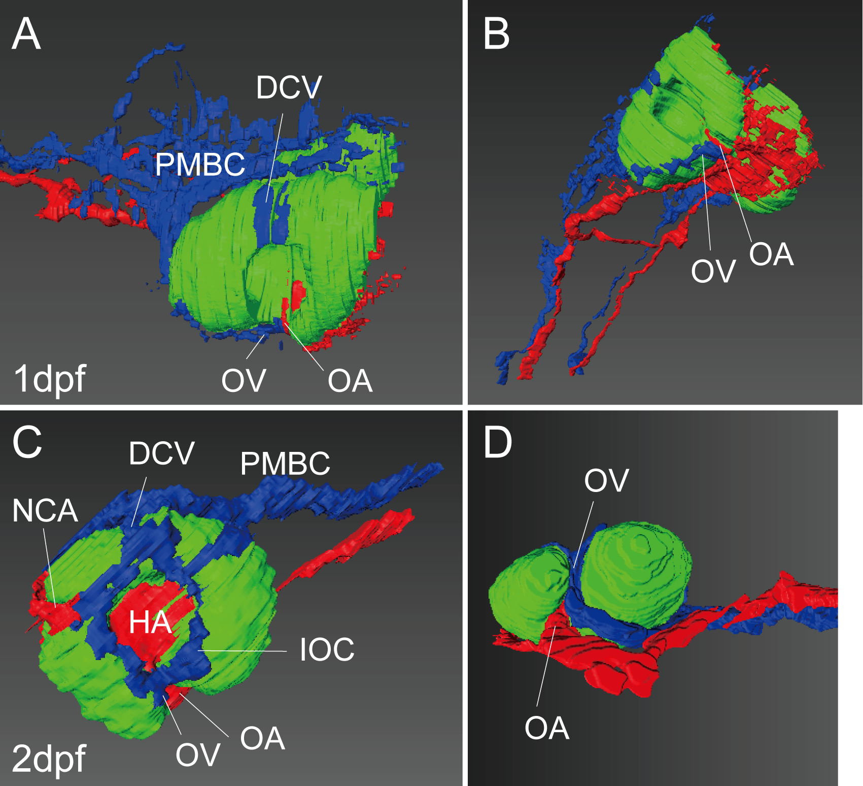
Fig. 10
Three-dimensional reconstruction of the ocular region.
Reconstructed images of the serial sections of GMA-embedded (A and B) and TECHNOVIT 7100-embedded (C and D) WT zebrafish embryos at 1(A and B) and 2(C and D) dpf. Lateral (A and C), ventral-lateral (B) and ventral (D) views. The artery, vein, and optic vesicle are colored in red, blue, and green, respectively.
Acknowledgments
This image is the copyrighted work of the attributed author or publisher, and
ZFIN has permission only to display this image to its users.
Additional permissions should be obtained from the applicable author or publisher of the image.
Full text @ PLoS One