
|
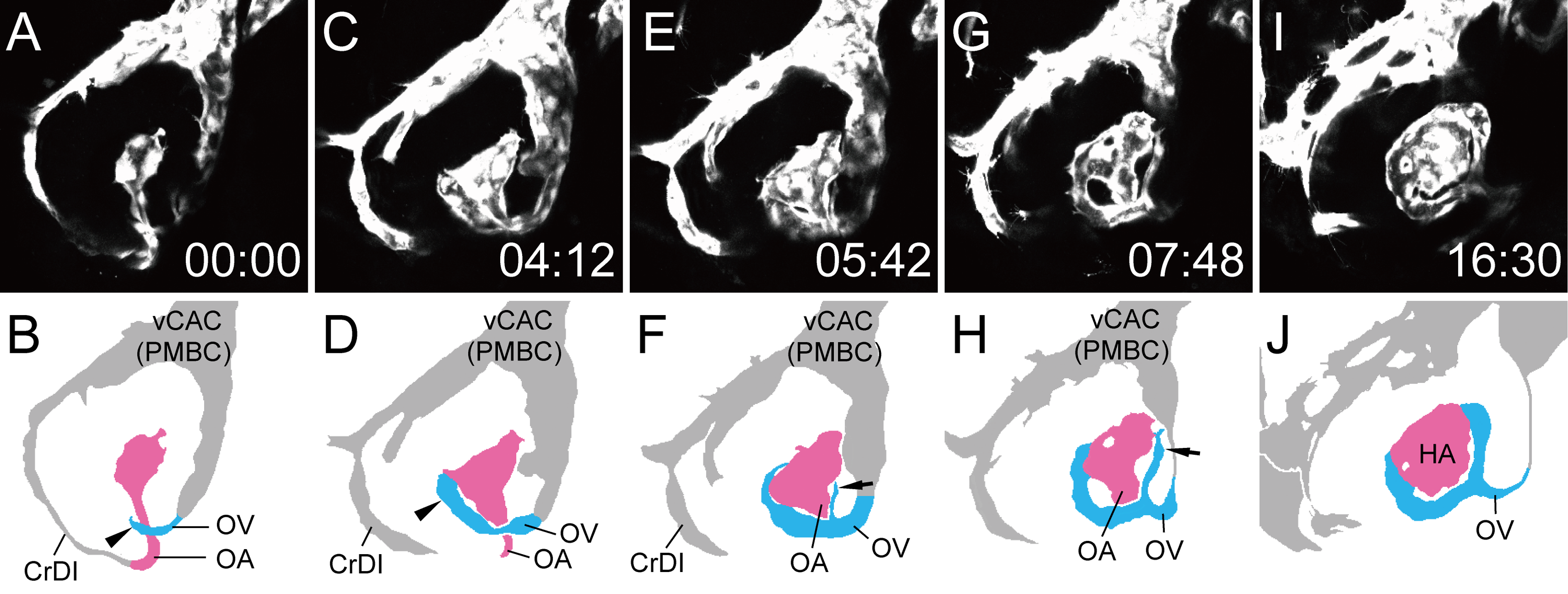
Fig. 6
Circular vessel formation connecting the OA with the OV.
Selected time-lapse images of a living Tg(flk1:EGFP)k7 embryo from 1 dpf (S4 Movie) (A, C, E, G and I) and their schematic diagrams (B, D, F, H and J). The time (hours:minutes) from the first frame is labeled in each image (A, C, E, G and I). Rostral is facing left and dorsal is facing upward. The formation of the left hyaloid vasculature was mainly observed. To visualize the formation of circular vessel connecting the OA with the OV, only the selected slices from S3 Movie were projected. Ocular vessels in the schematic diagrams are colored (OA: pink, and OV: sky blue). Arrowheads in B and D indicate the rostral sprout from the OV. Arrows in F and H indicate the caudal sprout from the OV.