
Fig. S2
- ID
- ZDB-IMAGE-111018-10
- Publication
- Patra et al., 2011 - Nephronectin regulates atrioventricular canal differentiation via Bmp4-Has2 signaling in zebrafish
- All Figures
- Figures for Patra et al., 2011
|
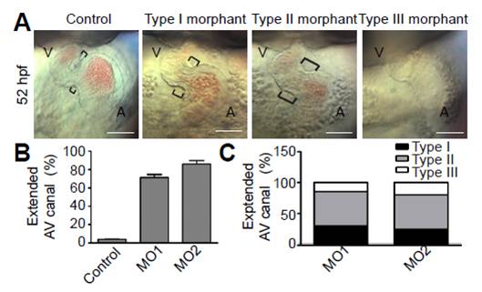
Fig. S2 Classification of AV canal defects. (A) Nomarski images at 52 hpf representing AV canal extension defects. Depending on severity, phenotypes were classified as type I (mild extension of the AV canal), type II (obvious extension of the AV canal) and type III (straight heart). Brackets indicate the AV boundary. Scale bars: 50 μm. (B) Quantitative analysis of embryos in terms of AV canal extension in total (n>150 from three independent experiments; mean ± s.e.m.). (C) Quantitative analysis in terms of type I, type II and type III AV canal extension. Note that type II was the most common phenotype (over 50%) in both MO1- and MO2-injected embryos.