
Fig. 1
- ID
- ZDB-IMAGE-110816-36
- Publication
- Zhao et al., 2011 - Analysis of MicroRNA Expression in Embryonic Developmental Toxicity Induced by MC-RR
- All Figures
- Figures for Zhao et al., 2011
|
Fig. 1
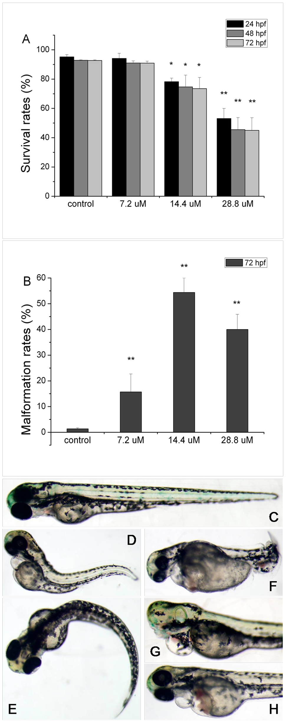
The survival, malformation rates and different developmental defects of zebrafish embryos induced by MC-RR.
(A) The survival rates of embryos after MC-RR exposure at 24, 48 and 72 hpf, respectively. (B) The malformation rates of embryos after MC-RR treatment at 72 hpf. Various morphological deformities were detected in MC-RR-treated embryos, including bent tail/body axes (D, E, F), cyclopia (G), as well as edema in pericardial sac (PS) and hatching gland (HG) (H), compared to control embryos (C). Values are expressed as mean ± SD (*indicates significant change compared to control at p<0.05, **indicates significant change compared to control at p<0.01).