FIGURE

Fig. 3

ID
ZDB-FIG-240308-6
Publication
Koparir et al., 2024 - Zebrafish as a model to investigate a biallelic gain-of-function variant in MSGN1, associated with a novel skeletal dysplasia syndrome
Other Figures
All Figure Page
Back to All Figure Page
Fig. 3

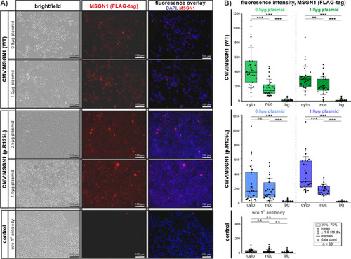
In vitro transfection of HEK 293T cells with CMV:MSGN1 (WT)-FLAG-tag and CMV:MSGN1 p.(Arg125Leu)-FLAG-tag plasmids at different concentrations. A Immunofluorescence showing expression of tagged MSGN1 proteins (WT) or p.(Arg125Leu) after 48h of transfection. Corresponding experimental controls are shown in Additional file 4: Fig. S3A. B Quantification of MSGN1 localization in transfected cells. Fluorescence signals in a by single z-plane were visualized by confocal laser-scanning microscopy and subsequently signal intensity was measured in single cells (cyto: cytoplasm; nuc: nucleus) and outside of cells (bg: background). Graphs show signal intensity measurements of the RFP channel (MSGN1 Flag-Tag) of 30 cells per experimental group and 30 background positions. Signal intensity measurements of the corresponding DAPI channel for nucleus identification are given in Additional file 4: Fig S3B. Values are given in Additional file 1: Excel file S1

Expression Data

Expression Detail
Antibody Labeling
Phenotype Data

Phenotype Detail
Acknowledgments
This image is the copyrighted work of the attributed author or publisher, and ZFIN has permission only to display this image to its users. Additional permissions should be obtained from the applicable author or publisher of the image. Full text @ Hum. Genomics