
Fig. 3
- ID
- ZDB-FIG-160714-13
- Publication
- Kawauchi et al., 2016 - Using mouse and zebrafish models to understand the etiology of developmental defects in Cornelia de Lange Syndrome
- Other Figures
- All Figure Page
- Back to All Figure Page
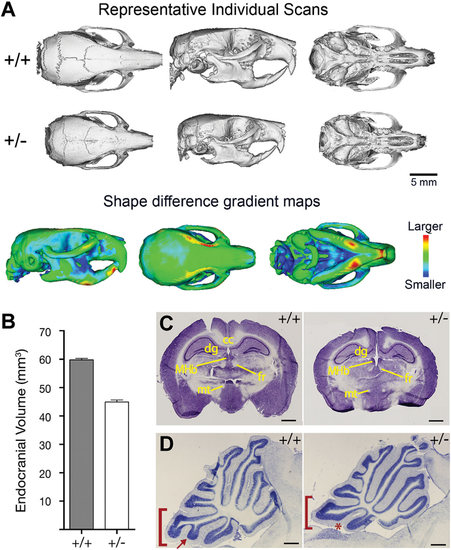
Craniofacial and neuroanatomical abnormalities in Nipbl+/- mice. (A) Top two panels show representative reconstructions of individual scans based on micro-CT analysis of wildtype (top panel) and Nipbl+/- (middle panel) adult mice. Dorsal, lateral, and ventral views are shown. Bottom panel shows shape distance heat maps of wildtype overlaid with mutant skulls, from the entire dataset. The degree of shape change in specific bones is shown by different colors, as indicated in the heat map at right. (B) Endocranial volume (measured from CT scans) is reduced by 25% in in Nipbl+/- mice compared to wildtype. (C) Nissl-stained coronal sections of wildtype (+/+) and Nipbl-deficient (+/-) adult brains. MHb, medial habenular nucleus; fr, fasiculus retroflexus; mt, mammillothalamic tract; dg, dentate gyrus; cc, corpus callosum. Scale bar =1 mm. (D) Cerebellar vermal hypoplasia in Nipbl+/- mice. Folium IX (bracket) and its ventral subfolium (arrow, asterisk) are affected. Scale bar = 500 µm. Adapted from [Kawauchi et al., 2009]. |